Cardiac Surgical Procedures
SURGICAL PROCEDURES
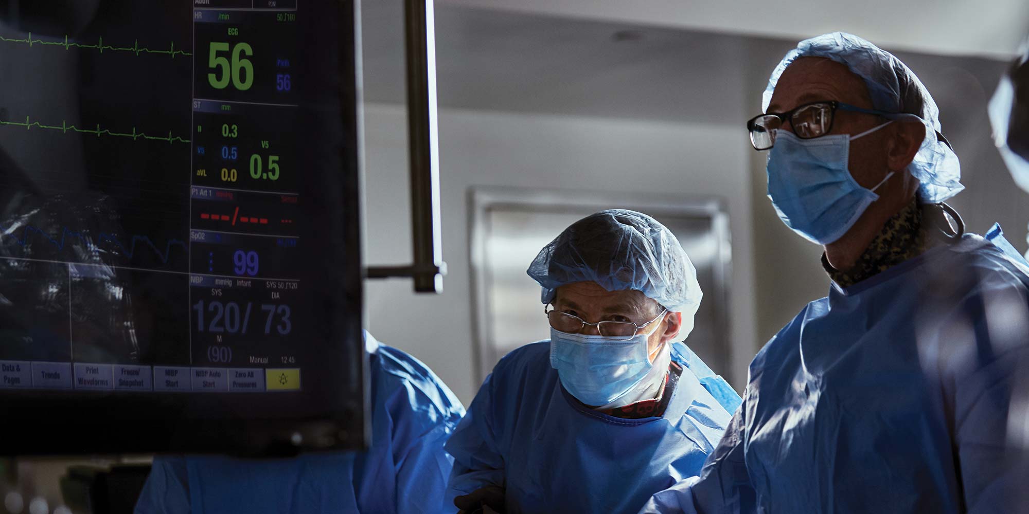
Performing the first open heart surgery in 1958, St. Joseph's Hospital Health Center is a leader in cardiac care. A pioneer in prevention, detection and the treatment of heart disease, St. Joseph’s performs more than 900 open heart procedures per year. Achieving excellence in cardiac surgery is truly a team effort. The cardiac surgery team includes the cardiac surgeon, cardiac anesthesiologist, physician assistants, nurse practitioners, perfusionists (those who run the heart-lung machine), surgical techs, OR, ICU and step-down unit nurses, physical therapists, social workers and case managers. Listed below are some highlights of the St. Joseph’s cardiac surgical program.
- Consistent clinical excellence, as evidenced by excellent results in the New York State Department of Health database over a 30-year period.
- Top five among all New York State cardiac surgical programs in terms of overall volume based on recent 2012 and projected 2013 volume data.
- Three star-rated program (3 out of 3 stars) from the Society of Thoracic Surgeons (STS) for overall quality, which places St. Joseph’s in top 5-10 percent of all programs in the United States.
- The Society of Thoracic Surgeons (STS) database is the largest clinical database in the world and the three star rating defines clinical excellence in cardiac surgery.
- Two cardiac surgeons (Dr. Zhou and Dr. Lutz) trained in robotic surgery with extensive clinical experience in robotic-assisted coronary artery bypass and mitral valve repair.
- Drs. Zhou and Lutz are also certified robotic cardiac surgery proctors (trainers) and have trained many surgeons and programs through the northeast in robotic techniques, including the Cleveland Clinic.
- Entire cardiac surgical staff extensively trained in minimally invasive procedures.
Procedures
Coronary Artery Surgery
Coronary Artery Bypass Grafting (CABG)
Coronary artery bypass grafting (CABG), also called heart bypass, bypass, or open heart surgery, represents the surgical treatment of coronary artery disease (CAD). It is important to recognize that CAD is a systemic disease with a genetic component that even with surgery or stenting requires medical treatment and risk-factor modification.
Basic medical treatment for CAD includes an anti-platelet agent (Aspirin) Beta-blocker medication, and a statin medication (anti-cholesterol agent). The most beneficial risk-factor modification for patients is to quit smoking followed by consistent exercise and proper diet. CAD can be treated with medical therapy, percutaneous coronary intervention (PCI or stenting), or CABG surgery. The goal is to relieve angina (chest pain or discomfort that may occur if an area of your heart muscle doesn’t get enough oxygen-rich blood) and reduce the risk of death from coronary artery disease. While PCI has become more common (and is less invasive) than CABG as a treatment of CAD, recent studies suggest that CABG offers a better long-term outcome (reduced death rate) compared to PCI. This is especially true in diabetic patients. Further studies will help define the best treatment option for patients.
During the CABG procedure, arteries (most commonly the left internal mammary artery [LIMA]) or veins (saphenous vein from the leg) from elsewhere in the patient's body are grafted (moved from one part of the body to another) to the coronary arteries to bypass atherosclerotic narrowing (thickening of the artery wall as a result of the accumulation of fatty materials such as cholesterol) and improve the blood supply to the heart muscle. It is standard of care in coronary bypass surgery to use at least one internal mammary artery. Newer data suggests that using both internal mammary arteries is beneficial and improves long-term survival and durability of the CABG procedure. St. Joseph's surgeons are very aggressive in using both internal mammary arteries to achieve the most benefit for the patient. CABG surgery is usually performed with the heart stopped, which requires placing the patient on the heart-lung machine or cardiopulmonary bypass.
Minimally Invasive Coronary Artery Bypass Grafting (CABG) Options
Off-pump Coronary Artery Bypass Grafting (OPCAB)
Off-pump coronary artery bypass (OPCAB) is a technique of performing bypass surgery without the use of cardiopulmonary bypass (the heart-lung machine). While traditional CABG is the gold standard, recent evidence suggests that the OPCAB technique may offer benefit for selected patients including women and high-risk patients, as well as patients who are not candidates for a traditional CABG procedure due to extensive aortic disease.
Robotic-assisted Coronary Bypass Grafting
The da Vinci® Surgical System, or da Vinci® robot as it is commonly called, is a $2.3 million device that allows a variety of complex surgeries to be performed through small incisions. The da Vinci® robot facilitates this by greatly enhancing the surgeon’s visualization and instrument dexterity. Controlled remotely, the robot, in essence, places the surgeon’s eyes and hands at the surgical field. The surgeon sits at a console separate from the patient and views a 3D image of the surgical field while controlling instruments that mimic the complex motions of the human hand. The surgeon’s technique is actually enhanced by the da Vinci® robot’s ability to scale motion and filter any tremor, thereby allowing very precise movements. The da Vinci® robot never functions independently; it is completely controlled by the surgeon.
In a robotic-assisted coronary artery bypass procedure, the left internal mammary artery (LIMA) is dissected off the inside of the chest wall using the da Vinci® Surgical System and the pericardium (sac around heart) is opened to identify the target coronary artery. The LIMA is then connected to the target coronary artery through a small incision (mini-thoracotomy) between the ribs. A one or two vessel bypass is possible through this approach, which is also called minimally invasive direct coronary artery bypass surgery (MIDCAB).
Since robotic surgery is performed through small incisions, the time to full recovery is much shorter. Conventional cardiac surgery is performed through a sternotomy (or breast bone dividing) incision, which limits the patient’s activity for two to three months as they recover. With robotic surgery there is no broken bone to heal so most patients are fully recovered within one month. For most patients this translates into a faster return to work and other activities such as running, biking and golfing. The patient’s length of time in the hospital is also shorter, averaging three to four days as opposed to six or seven days with conventional cardiac surgery.
For more information on other da Vinci® cardiothoracic procedures, click here.
Hybrid Surgery: Combining Robotic-assisted Coronary Bypass with Stenting of Other Coronary Arteries
For patients with three-vessel coronary artery disease who want a minimally invasive approach, a hybrid approach is available and is becoming more popular. This approach involves the cardiac surgeon and cardiologist working as a team to treat the patient’s coronary artery disease in the most minimally invasive way possible. The cardiac surgeon performs a robotic-assisted single-vessel coronary bypass and the cardiologist places a stent (a hollow mesh tube used to prop open a narrowed coronary vessel) to treat the disease in the other coronary arteries. St. Joseph’s hospital is one of the few centers in Upstate New York that offers this procedure.
Endoscopic Saphenous Vein Harvest
Traditionally, the saphenous vein was dissected or harvested from the leg through a large open incision. The endoscopic approach is a minimally invasive approach in which surgeons use an endoscope (thin surgical tube with a light, camera, and instrument on the end) to dissect the vein carefully through a small incision below the knee. This approach has been used for many years at St. Joseph’s and is currently the industry standard for grafting (moving arteries or veins from one part of the body to another).
Aortic Valve Surgery
The aortic valve separates the left ventricle from the aorta (the largest artery in the body), has three thin leaflets, and is the final valve blood flows through as it exits the heart. When diseased, the aortic valve can become either stenotic (tight) or insufficient (leak). Aortic valve disease can be either congenital (present at birth) or acquired (develops over time). The most common congenital problem is a bicuspid (two leaflet) aortic valve and the most common acquired problem is called calcific aortic stenosis. In the past rheumatic fever was a major cause of aortic valve disease, but is less common today.
Once the aortic valve becomes severely diseased and the patient develops symptoms (commonly angina, syncope [passing out], and shortness of breath) surgery is indicated and beneficial compared to the alternative of observation. It is important to note that unlike coronary artery disease there is no effective medical treatment of severe aortic valve disease. It is also important to note that the trend in the medical literature is toward earlier surgery. For patients with aortic valve stenosis (narrowing of aortic valve), the valve is generally heavily calcified and aortic valve repair is not possible, and as such the valve has to be replaced. For patients with aortic valve insufficiency, aortic valve repair is possible in certain patients, but overall replacement is more common. The surgical options are listed below.
Aortic Valve Replacement (AVR)
Aortic valve replacement involves removing the patient’s diseased valve and replacing it with one of the options below. Conventional replacement involves placing the patient on the heart lung machine.
Bioprosthetic Valve (Tissue: pig or cow)
The bioprosthetic valve is also referred to as a tissue valve and most commonly comes from a pig or cow. The major advantage for the patient is that they don’t have to take long-term Coumadin (a blood thinner). The major disadvantage is that tissue valves are not as durable as mechanical valves. The bottom line is that since cardiac surgery itself is relatively young (40 years essentially), we don’t have true long-term data on any of the major recent advances. Ten to 15 year data on the latest generation tissue valves show the reoperation rate can be estimated at 10 percent at 10 years. About 80 percent of all valves replaced in the United States today are replaced with tissue valves.
Mechanical Valve
Mechanical valves have the advantage of being very structurally durable with good hemodynamic (blood movement) properties. The major disadvantage for the patient is the need to take long-term Coumadin (a blood thinner, also called Warfarin), which carries a small constant increased risk of bleeding. The guidelines recommend a mechanical valve for any patient under 60, however the most recent guidelines give very strong consideration for the patient’s lifestyle preference and willingness to take long-term Coumadin. The trade-off for not choosing a mechanical valve and being off Coumadin is an increased risk of re-operation in the future.
Minimally Invasive Approach: Mini-sternotomy, Mini-thoracotomy & TAVR
A minimally invasive approach is an option for patient’s requiring aortic valve replacement and is very commonly performed at St. Joseph’s. There are two basic approaches, either a mini-thoracotomy (small incision between the ribs) or a mini-sternotomy (small upper partial breastbone dividing incision) approach. In general, both approaches allow the patients to get back to full activity faster compared to a conventional sternotomy approach.
For patients who are not candidates for a conventional or minimally invasive AVR there is now an option for patients. This option is called Transfemoral Aortic Valve Replacement (TAVR) and represents hope for a group of patients for whom and until recently there was no treatment option. TAVR involves the cardiologist and cardiac surgeon working as a team to place a new valve via a catheter placed in the femoral artery (groin artery). The only incision for the patient is a small groin incision. This approach is very new and currently is reserved for patients who are not candidates for a conventional AVR. It is anticipated that this procedure will expand in the future. For more information on TAVR, click here.
Aortic Valve Repair
In certain patients it is possible to repair the aortic valve. This is typically in patients who have an aortic aneurysm (ballooning of the artery) and the aortic valve leaflets are normal. The Yacoub and David Procedure are the two aortic valve repair procedures that are performed at St. Joseph’s. In both procedures the aortic wall is replaced and the aortic valve is spared, leaving the patient with his or her own valve. When functioning normally, the patient’s own valve is always superior to any replacement valve.
Mitral Valve Surgery
The mitral valve is located between the left atrium and left ventricle and essentially separates the heart from the lungs. The valve is a thin parachute like structure that is composed of two leaflets. The leaflets are attached to the ventricle by thin cords and when the heart contracts the leaflets seal and blood is forced out the aortic valve. The valve was named mitral by the early anatomists because it resembles a bishop’s mitre (hat). The mitral valve can become diseased either due to a leaflet (organic) or ventricular (functional) problem. The most common mitral valve problem is mitral insufficiency (leakage) caused by mitral valve prolapse. Mitral insufficiency is also commonly caused by a cardiomyopathy (weakening of the heart muscle). Mitral valve stenosis (narrowing) is caused mainly by rheumatic heart disease and is much less common today. When mitral insufficiency or stenosis is severe and the patient develops symptoms (shortness of breath and fatigue are the most common) then mitral valve surgery is indicated and preferable to medical treatment. Recent studies also suggest that patients with severe mitral insufficiency and no symptoms also benefit from surgery if the valve can be repaired. The guidelines also imply that because mitral valve surgery is generally elective, patients should seek out surgeons and centers experienced in mitral valve repair.
Mitral Valve Replacement
In some patients due to a severely diseased mitral valve (most commonly rheumatic pathology) a repair is not possible and the valve has to be replaced.
Bioprosthetic Valve (Tissue: pig or cow)
A bioprosthetic valve offers the benefit of not having to take long-term Coumadin (blood thinner) with the downside being a less durable option compared to a mechanical valve. Recent studies estimate the chance of a re-operation with a bioprosthetic mitral valve at approximately 15 percent at 10 years.
Mechanical Valve
A mechanical mitral valve is a very durable option for the patient with an extremely small chance of structural failure. The downside for the patient is the requirement to take life-long Coumadin (blood thinner). Given this substantial lifestyle restriction, most patients will choose a bioprosthetic valve.
Mitral Valve Repair
Most mitral valve pathology today is either mitral valve prolapse or a dilated (stretched) mitral valve due to a cardiomyopathy (disease of the heart muscle) and is repairable in most cases. If the valve CAN be repaired, repair is preferable to replacement and offers the patient the benefits of enhanced short and long-term survival, improved quality of life, better heart function and freedom from blood thinners.
Robotic-assisted Mitral Valve Repair
Patients with isolated mitral valve disease that can be repaired are candidates for a robotic-assisted approach. For the surgeon, the da Vinci® Surgical System provides enhanced visualization of the valve and increased instrument dexterity, thereby facilitating a precise repair. For the patient, the port approach via the right side of the chest avoids the breastbone dividing incision and means a much faster return to full activity and shortened hospital stay.
Tricuspid Valve Surgery
The tricuspid valve is located between the right atrium and the right ventricle in the heart. When heart valves become damaged or diseased, they may not function properly. Conditions which may cause heart valve dysfunction are valvular stenosis (stiffening) and valvular insufficiency (regurgitation). The tricuspid valve most commonly becomes diseased secondary to another valve problem (most commonly mitral) or because of infection. When the tricuspid valve becomes severely diseased and the patient develops symptoms (most commonly edema [fluid build-up] or fatigue) then surgery is indicated and beneficial compared to medical treatment. The options are listed below.
Tricuspid Valve Replacement
In some cases when the valve can’t be repaired (most commonly infection) replacement is the only option. This is most commonly done with a bioprosthetic or tissue valve (pig or cow).
Tricuspid Valve Repair
Most tricuspid valve disease is secondary to other valve problems and can be repaired. In general and if feasible, repair is preferable to replacement. When diseased, the tricuspid becomes dilated (stretches like an old shoe) and the thin leaflets that normally seal and prevent blood from leaking through the valve don’t meet. This results in blood flowing in the wrong direction. The most common repair technique is called an annuloplasty. This involves placing a dacron band around the tricuspid valve to reduce the diameter of the valve to a normal size. This results in the leaflets coapting (meeting) properly and corrects the leak.
Minimally Invasive Options
A tricuspid valve repair or replacement can be (and is commonly performed at St. Joseph's) through a minimally invasive approach either with or without the da Vinci® Surgical System. This involves making a small incision between the ribs on the right side of the chest called a mini-thoracotomy. In selected cases the surgeon may choose to use da Vinci® to assist in this approach. Like most minimally invasive valve procedures, this procedure requires placing the patient on the heart-lung machine which is typically done by accessing the femoral artery and vein through a small groin incision.
Congenital Heart Procedures
Atrial Septal Defect (ASD) Repairs
An ASD is a heart defect existing at birth where a hole is present in the atrial septum – the dividing wall between the two upper chambers of the heart known as the right and left atria. Most large ASDs are repaired by congenital heart surgeons when the patient is a child, but occasionally adults are diagnosed with an ASD. Most are repaired if the patient has associated symptoms or if the defect is found when the patient is undergoing surgery for another cardiac problem. Very small ASDs may not need treatment.
Ventricular Septal Defect (VSD) Repairs
A VSD is a hole between the two main pumping chambers (ventricles) that is normally repaired by congenital heart surgeons when the patient is very young. Occasionally a VSD can be caused by a massive heart attack and emergent repair is generally required.
Surgical Myomectomy
A surgical myomectomy is performed for asymmetric septal hypertrophy (also known as hypertrophic cardiomyopathy) or when the heart muscle becomes thick. This genetic condition can result in sudden cardiac death and is characterized by abnormal thickening of the ventricular septal muscle below the aortic valve. In its severe form, it is difficult for blood to leave the heart and is often associated with symptoms such as shortness of breath or syncope (passing out). Initial treatment is medical, but the definitive treatment is surgical myomectomy in which a segment of left ventricular muscle is excised and the outflow tract is widened.
Cardiac Tumor Excision
Tumors in the heart are rare. When they do occur, they may be either malignant (have the ability to spread to other sites in the body or to invade and destroy tissues) or benign (not cancerous).
Most cardiac tumors are benign, but it important to note that even benign tumors can be life-threatening. Often these tumors are diagnosed when the patient experiences a neurologic event such as a transient ischemic attack (TIA) or a "mini" stroke. Benign tumors include myxoma (most common type), rhabdomyomas and fibromas. Malignant heart tumors occur most often in children. These include angioscarcomas, fibroscarcomas and rhabdomyosarcomas.
Malignant tumors that occur elsewhere in the body and spread to the heart are more common than those that originate in the heart. Excision is surgical removal of the tumor and in most cases is the treatment of choice. Depending on the type of tumor, treatment may be combined with radiation and chemotherapy.
Congestive Heart Failure (CHF) Surgery
Congestive heart failure (CHF) is becoming an increasingly common form of heart disease. Like coronary artery disease, in patients with CHF medical therapy and risk-factor modification is extremely important. Basic medical therapy often includes a beta blocker, ACE inhibitor and diuretic medications. Risk-factor modifications include quitting smoking, as well as proper diet and exercise. For patients with severe CHF and symptoms surgical treatment is often indicated and beneficial.
Ventricular Assist Device (VAD)
A Ventricular Assist Device (VAD) is essentially an artificial heart pump that takes over the basic heart pumping function in patients with severe congestive heart failure (CHF). A VAD can be temporary or permanent. St. Joseph’s Hospital occasionally places the temporary centrimag VAD for patients with severe CHF. Permanent VADs, such as the Heartmate II VAD, are generally placed by heart transplant surgeons at designated heart transplant centers.
Extracorporeal Membrane Oxygenation (ECMO)
Extracorporeal membrane oxygenation (ECMO) is essentially an artificial heart and lung and is occasionally placed at St. Joseph’s for patients with severe heart and lung failure or isolated lung failure, as occasionally occurs in patients with severe influenza. ECMO is also used for respiratory failure.
Minimally Invasive Left Ventricular Pacemaker Lead Insertion in Conjunction with Biventricular Pacing
In some patients with congestive heart failure (CHF), the timing of pumping between the ventricles is not synchronized (i.e., the right and left ventricles do not pump at the same time). In this group of patients, implanting a biventricular pacemaker that paces both ventricles simultaneously can improve a patients quality of life and survival. This is most commonly performed with a catheter approach, but occasionally the left ventricular lead can’t be placed and needs to be placed by a cardiac surgeon through a minimally invasive approach. St. Joseph’s is a regional leader in this procedure.
Aneurysm Surgery
A thoracic aortic aneurysm is a bulging, weakened area in the wall of the aorta (the largest artery in the body), resulting in an abnormal widening or ballooning greater than 50 percent of the vessel's normal diameter. The thoracic aorta is located in the chest area and is the major blood vessel taking blood away from the heart and toward the other organs. An aortic aneurysm can be caused by a combination of many factors, including high blood pressure, smoking, and genetic factors such as bicuspid aortic valve (an aortic valve with only two leaflets instead of three) and Marfan’s syndrome (a genetic disorder of the connective tissue). Aortic aneurysms are often diagnosed incidentally when the patient undergoes a chest xray or CT scan for another problem. Once diagnosed, it is important that the patient be followed by a cardiac surgeon.
Repair of Ascending, Arch, or Descending Aortic Aneurysm
Ascending and arch aortic aneurysms are repaired by cardiac surgeons and generally require an open conventional repair. Descending aortic aneurysms can often be repaired using endovascular techniques (stent grafts) and are performed by cardiac and vascular surgeons.
Repair of Aortic Coarctation
Aortic coarctation is a narrowing in the first part of the descending aorta and is often diagnosed in childhood and repaired by congenital heart surgeons. Occasionally, patients are diagnosed as adults and repaired by an adult cardiac surgeon. An endovascular approach (minimally invasive surgical approach to enter the body via major blood vessels) may be an option in some cases.
Repair of Aortic Dissection
Aortic dissection is a tearing within the wall of the aorta (the largest artery in the body), and represents a life-threatening emergency requiring emergent medical and surgical treatment. Dissections are often associated with uncontrolled high blood pressure or smoking and may occur in a patient with an aneurysm (bulging of a blood vessel). Aortic dissections are classified into two types: Type A and Type B dissections. Type A dissections start in the ascending aorta and require emergent surgical repair to prevent death from aortic rupture. Type B aortic dissections start in the descending aorta and can generally be managed medically with blood pressure control. Surgery in this group is reserved for those patients in whom medical management fails or have evidence of rupture.
Ventricular Aneurysm Repair (Aneurysmectomy)
A ventricular aneurysmectomy is a surgical procedure performed to repair an aneurysm (a bulging, weakened area in the wall of a blood vessel resulting in an abnormal widening or ballooning greater than 50 percent of the vessel's normal diameter) in the ventricle (a fluid filled cavity in the heart). Ventricular aneurysms are one of the many complications that may occur after a heart attack. They usually arise from a patch of weakened tissue in a ventricular wall, which swells into a bubble filled with blood. This, in turn, may block the passageways leading out of the heart, leading to severely constricted blood flow to the body.
Atrial Fibrillation Surgery
Atrial fibrillation (AF) is a disorganized rhythm of the atria (top heart chamber) that results in a decrease in cardiac output (amount of blood the heart pumps over time). AF can be either intermittent (comes and goes) or chronic (all the time). Patients with AF often have a fast heart rate and can develop symptoms such as shortness of breath and palpitations. Patients with AF can often be treated successfully with medications that slow the heart rate and help convert the rhythm to normal sinus rhythm. In addition, patients with AF almost always need to take a blood thinning medication such as Coumadin to reduce the risk of stroke associated with AF.
For patients in whom medical therapy is not successful and have symptoms, catheter or surgical ablation may be an option. Catheter ablation is performed by an electrophysiologist and involves the cardiologist passing a catheter (a flexible tube) into the heart and creating scar tissue to separate the source of atrial fibrillation from the heart. Catheter ablation is less invasive than surgical ablation, but has a higher recurrence rate.
The surgical option is called a MAZE procedure and was first described in the 1980s by cardiac surgeon Dr. Jim Cox. The MAZE procedure involves creating scars in a specific pattern in the right and left atria. The result is that the electrical cardiac signal is forced in an orderly pathway and the patient experiences normal sinus rhythm. The original MAZE procedure was performed by cutting and sewing the atrial tissue in a specific pattern. It was very invasive and was only performed by a few surgeons. Over the past 10 years, newer energy sources and techniques have been developed that have reduced the invasiveness of the MAZE procedure and the procedure is very commonly performed.
The MAZE procedure can be performed in conjunction with another procedures or as a stand-alone procedure for patients with symptomatic AF. It is important to note that it takes about six months following the procedure to know for sure if the MAZE procedure has been successful. True success is defined as the patient being in normal sinus rhythm off of all medications.
Stand-alone Options
Galaxy Procedure
The Galaxy Procedure is a minimally invasive option for atrial fibrillation (AF) in which scars are created on the outside of the left atrium by small radiofrequency energy catheters. As part of this procedure the left atrial appendage (a sac like structure that can contain blood clots) is bound to reduce the patient’s stroke risk. This procedure is performed through very small incisions in the patient’s right and left chest and is done without placing the patient on the heart-lung machine or stopping the heart. This procedure can also be performed as part of a hybrid procedure in which the cardiologist performs part of the procedure with a catheter.
Minimally Invasive Cryomaze Procedure
The Cryomaze Procedure is a minimally invasive option in which scars are created on the inside of the left and right atrium by a catheter (flexible tube) that freezes the atrial tissue. As part of this procedure the left atrial appendage (a sac like structure that can contain blood clots) is bound to reduce the patient’s stroke risk. This procedure is performed through a small incision in the patient’s right chest and is done on the heart-lung machine with the heart stopped.
In Conjunction with Another Procedure
The Galaxy or Cryomaze Procedure can also be performed when the patient has atrial fibrillation (AF) and is undergoing another cardiac surgical procedure (i.e., bypass or valve surgery). Both procedures are very safe, add very little time to the procedure, and are recommended to increase the patient’s chances of conversion to a normal sinus rhythm.

